In the paper
Unit 1 carries 2 of the 40 botany marks; with two chapters sharing them, this one averages one (MEC publishes weights by unit, not by chapter). The MEC scope line: Structure, types and biological role of proteins; Enzymes. Three ways it is asked:
- Recall: the bond joining amino acids; the class of lipase; who proposed induced fit; the vitamin behind NAD.
- Understanding: why extra substrate overcomes a competitive inhibitor; why boiling stops an enzyme for good but cold does not; why one amino-acid change sickles red cells.
- Application: reading Km from a rate figure; predicting what builds up when malonate is added; counting peptide bonds in a short chain.
Structure, types and biological role of proteins
Proteins are polymers of amino acids, containing C, H, O, N and usually S.
Amino acids and the peptide bond
- Proteins are built from 20 common amino acids. Each has a central alpha carbon carrying an amino group (–NH₂), a carboxyl group (–COOH), a hydrogen and a variable R group. Glycine's R group is a single hydrogen; cysteine's carries –SH.
- Plants make all twenty. Humans cannot make nine — the essential amino acids — and must eat them. Cereal protein is short of lysine and pulse protein of methionine, so rice with lentils (dal-bhat) supplies a better mix than either alone.
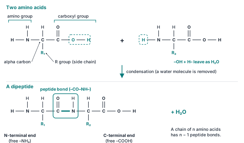
- Two amino acids join by a peptide bond (–CO–NH–) between the carboxyl of one and the amino group of the next, releasing one water molecule (condensation). A chain of n amino acids has n − 1 peptide bonds, a free N-terminal end and a free C-terminal end.
Figure 1 Peptide bond formation
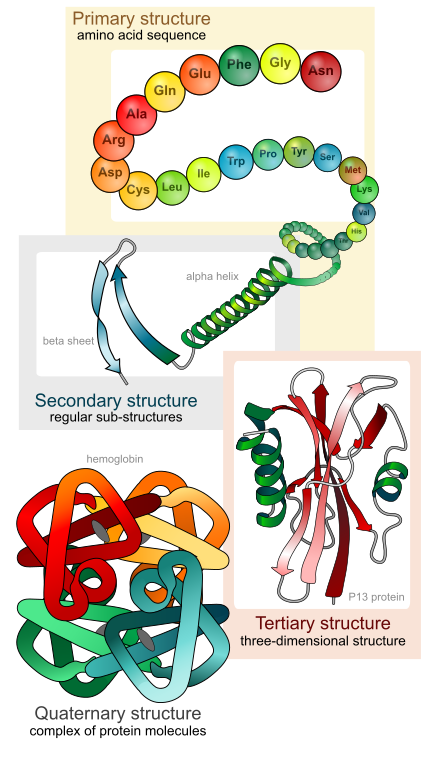
Four levels of structure
| Level | What it is | Held by | Example |
|---|---|---|---|
| Primary | Sequence of amino acids | Peptide bonds | Insulin: chain A of 21 and chain B of 30 amino acids, joined by two disulfide bridges |
| Secondary | Local coiling or pleating of the backbone | Hydrogen bonds between backbone C=O and N–H | Alpha-helix (3.6 residues per turn; keratin of hair); beta-pleated sheet (silk fibroin) |
| Tertiary | The whole chain folded into a 3-D shape | R-group interactions: hydrogen and ionic bonds, hydrophobic interactions, disulfide (S–S) bridges between cysteines | Myoglobin; most enzymes — the active site forms here |
| Quaternary | Two or more polypeptide subunits together | The same forces, between chains | Haemoglobin: two alpha + two beta chains |
Figure 2 The four levels of protein structure
Sequence dictates shape. In sickle-cell haemoglobin, valine replaces glutamic acid at position 6 of the beta chain; the altered molecules stick into fibres and bend red cells into sickles.
Denaturation. Heat, extreme pH, heavy-metal ions (Hg²⁺, Pb²⁺, Ag⁺) and organic solvents break the bonds of secondary, tertiary and quaternary structure. The protein unfolds and loses function, but its primary sequence and peptide bonds survive. Frying egg white is irreversible; mild denaturation can reverse when the agent is removed. Chaperone proteins help new chains fold correctly.
Types
| Fibrous | Globular | |
|---|---|---|
| Shape | Long parallel chains, often cross-linked | Compact, roughly spherical |
| In water | Insoluble | Soluble |
| Job | Structure | Enzymes, transport, hormones, antibodies |
| Examples | Keratin, collagen, silk fibroin | Enzymes, haemoglobin, insulin, albumin |
By composition:
- Simple proteins give only amino acids on hydrolysis: albumins, globulins (legumin of pea), glutelins (glutenin of wheat), prolamins (zein of maize, gliadin of wheat), histones (which pack DNA).
- Conjugated proteins carry a non-protein prosthetic group: nucleoproteins (chromatin, ribosomes), glycoproteins (mucin, membrane receptors), lipoproteins (membranes, blood transport), chromoproteins (haemoglobin, cytochromes, phytochrome), phosphoproteins (casein of milk), metalloproteins (ferritin with iron, plastocyanin with copper).
- Derived proteins are products of partial breakdown: proteoses, peptones, peptides.
Biological roles
| Role | Examples |
|---|---|
| Catalysis | Enzymes — amylase, RuBisCO |
| Structure | Collagen, keratin, tubulin |
| Transport | Haemoglobin (oxygen), membrane carriers and channels |
| Storage | Seed proteins in aleurone grains (zein, gluten), casein, ferritin (iron) |
| Hormones | Insulin, glucagon |
| Defence | Antibodies, fibrinogen, plant lectins |
| Movement | Actin, myosin; tubulin in the spindle |
| Sensing | Phytochrome (red and far-red light), hormone receptors |
Enzymes
Enzymes are biological catalysts. Holding substrates in the right orientation at the active site, they lower the activation energy, so the reaction reaches equilibrium sooner — but the equilibrium point itself does not move, and the enzyme comes out unchanged. Almost all are proteins; the exceptions are ribozymes (catalytic RNA, such as the peptidyl transferase of the ribosome).
| Year | Who | What |
|---|---|---|
| 1877 | Wilhelm Kühne | Coined the word "enzyme" ("in yeast") |
| 1894 | Emil Fischer | Lock-and-key model |
| 1897 | Eduard Buchner | Yeast extract ferments sugar with no living cells (zymase); Nobel 1907 |
| 1913 | Michaelis and Menten | Rate equation and Km |
| 1926 | James Sumner | Crystallised urease, proving an enzyme is a protein |
| 1958 | Daniel Koshland | Induced-fit model |
Parts of an enzyme
The protein part is the apoenzyme; the non-protein helper is the cofactor; together they make the active holoenzyme. Cofactors come in three kinds:
- Prosthetic group — organic, tightly bound: haem in catalase and peroxidase.
- Coenzyme — organic, loosely bound, shuttling hydrogen or groups between enzymes and regenerated after each cycle; most are built from B vitamins.
- Metal-ion activator — Zn²⁺ (carbonic anhydrase, DNA polymerase), Mg²⁺ (hexokinase and other kinases, RuBisCO), Mo (nitrogenase), Cu (cytochrome oxidase).
| Coenzyme | Vitamin | Carries |
|---|---|---|
| NAD⁺, NADP⁺ | Niacin (B3) | Hydrogen — dehydrogenases |
| FAD, FMN | Riboflavin (B2) | Hydrogen |
| Coenzyme A | Pantothenic acid (B5) | Acetyl groups |
| Thiamine pyrophosphate | Thiamine (B1) | Removal of CO₂ from pyruvate |
| Pyridoxal phosphate | Pyridoxine (B6) | Amino groups (transaminases) |
Naming and classification
Most names add -ase to the substrate (maltase, lipase, urease) or the reaction (dehydrogenase, polymerase); a few old names survive (pepsin, trypsin, ptyalin). The IUB (now IUBMB) Enzyme Commission gives each enzyme a four-part EC number. The Grade 11 text teaches six classes; a seventh, translocases, was added in August 2018.
| EC | Class | Reaction | Examples |
|---|---|---|---|
| 1 | Oxidoreductases | Oxidation–reduction (hydrogen or electron transfer) | Alcohol and succinate dehydrogenase, catalase, cytochrome oxidase |
| 2 | Transferases | Move a group other than hydrogen | Hexokinase (phosphate), transaminases (amino group) |
| 3 | Hydrolases | Break bonds by adding water | Amylase, lipase, pepsin, sucrase, urease |
| 4 | Lyases | Remove groups without water, leaving a double bond (or add to one) | Aldolase, carbonic anhydrase, pyruvate decarboxylase |
| 5 | Isomerases | Rearrange atoms within one molecule | Phosphoglucoisomerase, triose phosphate isomerase |
| 6 | Ligases | Join two molecules using ATP | DNA ligase, pyruvate carboxylase |
| 7 | Translocases | Move ions or molecules across membranes | ATP synthase, Na⁺/K⁺ pump |
How enzymes work
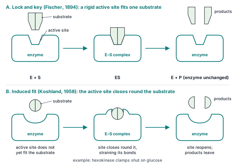
E + S ⇌ ES → E + P. In Fischer's lock-and-key model a rigid active site fits one substrate exactly, explaining specificity (urease acts on urea alone). In Koshland's induced fit, the active site is flexible and closes round the substrate as it binds, straining the bonds to be broken — hexokinase clamps shut on glucose.
Figure 3 Lock-and-key and induced-fit models
Factors affecting activity
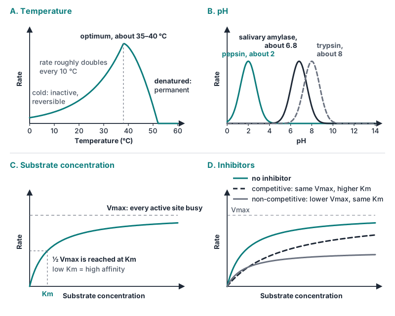
- Temperature: the rate roughly doubles or trebles for each 10 °C rise (Q10 about 2–3) up to an optimum (about 35–40 °C for most plant and animal enzymes); above it the protein denatures and activity collapses. Cold only inactivates, reversibly.
- pH: each enzyme has an optimum — pepsin about 2, salivary amylase about 6.8, trypsin about 8. Away from it the charges on active-site R groups change.
- Substrate concentration: rate rises with substrate, then levels at Vmax when every active site is busy (saturation). Km, the Michaelis constant, is the substrate concentration giving half Vmax; a low Km means high affinity.
- Enzyme concentration: with substrate in excess, rate is proportional to the amount of enzyme.
Inhibition
| Competitive | Non-competitive | |
|---|---|---|
| Resembles substrate? | Yes — a structural analogue | No |
| Binds | The active site | Elsewhere (an allosteric site) |
| Overcome by more substrate? | Yes | No |
| Vmax | Unchanged | Lowered |
| Apparent Km | Raised | Unchanged |
| Example | Malonate blocks succinate dehydrogenase (which turns succinate into fumarate); sulpha drugs mimic PABA in bacteria | Cyanide on cytochrome oxidase; Hg²⁺ and Ag⁺ binding –SH groups |
Figure 4 Enzyme rate against temperature, pH, substrate and inhibitors
Feedback (allosteric) inhibition: the end product of a pathway binds an allosteric site on the pathway's first enzyme and switches it off — isoleucine inhibits threonine deaminase. Allosteric activators do the reverse: ADP stimulates some glycolytic enzymes that ATP inhibits.
Numbers and names to memorise
| Item | Value |
|---|---|
| Amino acids in proteins | 20; nine essential for humans |
| Peptide bonds in a chain of n residues | n − 1 |
| Alpha-helix | 3.6 residues per turn; hydrogen bonds |
| Insulin | A chain 21, B chain 30 amino acids |
| Haemoglobin | Two alpha + two beta chains; sickle cell: Glu → Val at beta 6 |
| "Enzyme" / cell-free fermentation | Kühne 1877 / Buchner 1897 |
| First enzyme crystallised | Urease, Sumner 1926 |
| Lock and key / induced fit | Fischer 1894 / Koshland 1958 |
| Km | Substrate concentration at half Vmax |
| EC classes | Six in the Grade 11 text; translocases (EC 7) added 2018 |
| Optimum pH | Pepsin about 2, salivary amylase about 6.8, trypsin about 8 |
Traps
- Denaturation keeps the peptide bonds; hydrolysis breaks them.
- All coenzymes are cofactors, not all cofactors are coenzymes — a metal ion is a cofactor but not a coenzyme; a prosthetic group is bound tightly.
- Competitive raises Km and leaves Vmax; non-competitive lowers Vmax and leaves Km. Do not swap them.
- Low Km = high affinity, not low.
- Enzymes change the rate, not the equilibrium or the energy released.
- Cold inactivates, heat denatures.
- Class slips: carbonic anhydrase is a lyase, hexokinase a transferase, DNA ligase a ligase; hydrolases are EC 3.
- Not every enzyme is a protein: ribozymes are RNA.
Quick check
0 of 5 answered- 1The alpha-helix of a protein is held in shape mainly by:
- 2Adding a large excess of substrate restores an inhibited enzyme to its normal maximum rate. The inhibitor is most likely:
- 3Hexokinase moves a phosphate group from ATP to glucose. It belongs to the class:
- 4Enzyme P has a Km of 0.5 mM and enzyme Q a Km of 5 mM for the same substrate. At 0.5 mM substrate:
- 5A protein solution heated to 90 °C loses its activity. Which feature remains unchanged?
Sources
- OpenStax, Biology 2e, 3.4 Proteins and 6.5 Enzymes (consulted), read 2026-09-15.
- Wikipedia: Enzyme (Kühne 1877, Buchner, Sumner, Fischer 1894, Koshland), Enzyme Commission number, Translocase (EC 7, August 2018), Michaelis–Menten kinetics (Km, 1913), read 2026-09-15.
- NCERT Biology Class 11, ch. 9 Biomolecules; Nepal CDC Grade 11 Biology — consulted for the classification scheme, inhibition framing and terminology.
- Figures: credited in each caption (Wikimedia Commons and original diagrams).